How the Knee Functions During Walking: Mechanics, Movement, and Common Injuries
What does the knee do as you walk?
Function of the Knee
The knee is often considered a simple hinge joint, responsible for bending and straightening the leg. However, its mechanics are far more complex. The knee functions as a dynamic structure, accommodating movement across multiple planes while responding to forces from above (the pelvis and femur) and below (the tibia and foot). Understanding knee function requires looking at its three-dimensional movements and how they interact with surrounding structures.
Possible Mechanics of the Knee
Knee movement is dictated by the articulation between the femur, tibia, and meniscus. It primarily moves through two planes:
1. Sagittal Plane: This includes flexion (bending) and extension (straightening) of the knee.
2. Transverse Plane: This involves internal and external rotation of the knee, which is often overlooked but plays a crucial role in overall leg function.
The knee’s motion is largely influenced by the foot’s interaction with the ground. The tibia, which sits atop the talus, transmits movement from the foot to the rest of the body. When the foot pronates, the tibia internally rotates, influencing femoral rotation and, subsequently, knee mechanics.
Knee in Pronation
When the foot pronates, the talus moves medially, causing the tibia to internally rotate. This internal rotation of the tibia leads to increased internal rotation of the femur. However, because the medial tibial plateau is larger than the lateral, the femur has more room to rotate internally than the tibia. This results in an externally rotated knee joint, even though both the tibia and femur are internally rotating relative to the foot.
Key indicators of a knee in pronation include:
- A more pronated foot
- A foot turned outward
- An internally rotated talus
- A patella facing inward
- Reduced hip internal rotation
- Anterior pelvic tilt rotated away from the injured knee
When the foot is pronated, knee flexion is often observed. If the knee is hyperextended instead of flexed, it may indicate compensatory mechanisms that place excessive strain on the knee joint and its supporting structures.
Knee in Supination
Conversely, in a supinated foot, the tibia externally rotates, affecting knee alignment. If the foot is supinating properly, the knee can internally rotate to maintain proper mechanics. However, if the foot fails to supinate, the tibia and knee may be forced into excessive external rotation, reducing overall knee stability and limiting its ability to absorb shock effectively.
Key features of a knee in supination include:
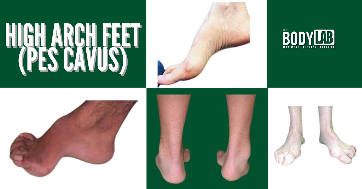
- A high-arched or rigid foot structure
- A foot with reduced medial ground contact
- Excessive external rotation of the tibia
- Increased stress on lateral knee structures
Improper knee positioning during supination can result in altered loading patterns, contributing to knee injuries over time.
Injuries of the Knee: ACL, PCL, LCL, MCL
The knee’s susceptibility to injury is largely dependent on its functional alignment and how forces are distributed through its structures. Here are some of the most common knee injuries and how they relate to movement mechanics:
Anterior Cruciate Ligament (ACL) Injuries
The ACL is commonly injured when excessive external rotation occurs in the knee, often due to poor foot mechanics and an inability to stabilise during dynamic movement. A pronated foot and internally rotated tibia increase the likelihood of ACL strain, particularly in sports requiring sudden directional changes.
Posterior Cruciate Ligament (PCL) Injuries
PCL injuries are typically caused by hyperextension or direct impact to the knee. If the knee remains in a hyperextended position due to poor foot posture, the PCL is subjected to undue stress, leading to instability and potential injury.
Lateral Collateral Ligament (LCL) Injuries
The LCL stabilises the outer knee and is often injured due to excessive lateral forces. When the foot fails to supinate properly, excessive strain is placed on the lateral knee, predisposing the LCL to damage.
Medial Collateral Ligament (MCL) and Meniscus Injuries
MCL injuries are common in individuals with excessive knee external rotation. Since the medial meniscus bears most of the weight in a pronated knee, excessive compression and shear forces can lead to meniscus tears. Identifying pronation-related knee stress can help prevent and rehabilitate MCL and meniscus injuries effectively.
Addressing Knee Injuries Through Postural and Movement Correction
The posture and resting alignment of the knee often dictate injury susceptibility. Rather than treating the injury in isolation, addressing the entire kinetic chain is crucial. Consider the following corrective strategies:
1. Encouraging foot supination to improve tibial positioning and reduce excessive external knee rotation.
2. Aligning the femur correctly by promoting external femoral rotation where necessary.
3. Encouraging proper knee extension without relying on compensatory hyperextension.
4. Adjusting pelvic alignment to ensure balanced weight distribution.
Many knee injuries are preventable if movement inefficiencies are identified and corrected early. Rather than seeing the knee as an isolated problem area, understanding its interaction with the foot and hip provides a more effective approach to treatment and injury prevention.
Understanding knee mechanics during walking involves examining its complex structure and movements.
How I Can Help
At The Body Lab, I specialise in assessing and improving movement mechanics to prevent and rehabilitate knee injuries. My approach integrates:
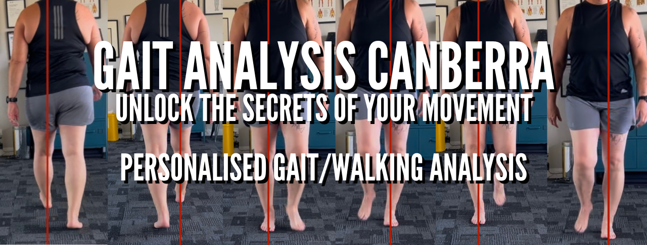
Gait and biomechanical analysis to identify dysfunctional movement patterns.
Hands-on therapy to restore proper alignment and function.
Individualised movement programs tailored to each client’s needs.
Workshops and educational sessions to help practitioners and individuals understand movement-based solutions for knee pain.
If you're struggling with knee pain or want to enhance your movement efficiency, book a consultation today and take the first step toward pain-free movement.
For further reading, consider these sources:
• Lower-limb walking pattern. (n.d.). Wikipedia. Retrieved from https://en.wikipedia.org/wiki/Lower-limb_walking_pattern
• Gait (human). (n.d.). Wikipedia. Retrieved from https://en.wikipedia.org/wiki/Gait_%28human%29
• Popliteus muscle. (n.d.). Wikipedia. Retrieved from https://en.wikipedia.org/wiki/Popliteus_muscle
These resources offer detailed insights into knee mechanics, gait patterns, and related injuries.
1. Neumann, D.A. (2016) Kinesiology of the Musculoskeletal System: Foundations for Rehabilitation. 3rd edn. St. Louis: Elsevier.
2. Escamilla, R.F. (2001) ‘Knee biomechanics of the dynamic squat exercise’, Medicine and Science in Sports and Exercise, 33(1), pp. 127–141. Available at: https://doi.org/10.1097/00005768-200101000-00020
3. Zaffagnini, S., Dejour, D. and Grassi, A. (2019) Knee Ligament Injuries: Extraarticular and Intra-articular Reconstruction Techniques. Berlin: Springer.
4. Griffin, T.M. and Guilak, F. (2005) ‘The role of mechanical loading in the onset and progression of osteoarthritis’, Exercise and Sport Sciences Reviews, 33(4), pp. 195–200. Available at: https://doi.org/10.1097/00003677-200510000-00009
5. Andriacchi, T.P. and Dyrby, C.O. (2005) ‘Interactions between kinematics and loading during walking for the normal and ACL deficient knee’, Journal of Biomechanics, 38(2), pp. 293–298. Available at: https://doi.org/10.1016/j.jbiomech.2004.02.010
6. Powers, C.M. (2010) ‘The influence of abnormal hip mechanics on knee injury: A biomechanical perspective’, Journal of Orthopaedic & Sports Physical Therapy, 40(2), pp. 42–51. Available at: https://doi.org/10.2519/jospt.2010.3337
7. Winter, D.A. (2009) Biomechanics and Motor Control of Human Movement. 4th edn. Hoboken: Wiley.
8. Besier, T.F., Fredericson, M., Gold, G.E., Beaupré, G.S. and Delp, S.L. (2009) ‘Knee muscle forces during walking and running in patellofemoral pain patients and pain-free individuals’, Journal of Biomechanics, 42(7), pp. 898–905. Available at: https://doi.org/10.1016/j.jbiomech.2009.01.032
9. Flandry, F. and Hommel, G. (2011) ‘Normal anatomy and biomechanics of the knee’, Sports Medicine and Arthroscopy Review, 19(2), pp. 82–92. Available at: https://doi.org/10.1097/JSA.0b013e31821b6c75
10. Della Croce, U., Camomilla, V., Leardini, A. and Cappozzo, A. (2005) ‘Human movement analysis using stereophotogrammetry. Part 4: Assessment of anatomical landmark misplacement and its effects on joint kinematics’, Gait & Posture, 21(2), pp. 226–237. Available at: https://doi.org/10.1016/j.gaitpost.2004.05.003