The High Arched Foot – What Are the Effects on the Body?
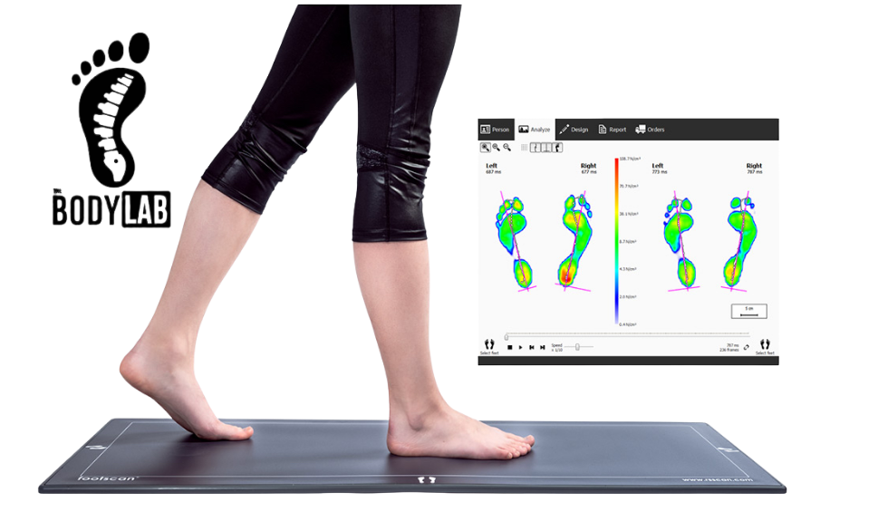
The High Arched Foot at The Body Lab
The structure of the foot is critical in supporting movement, posture, and balance. Among the various foot types, a high-arched foot (pes cavus) presents unique challenges that can impact movement, biomechanics, and long-term function. In this article, we will explore how foot arches influence overall movement, the differences between foot arch types, the effects of high-arched feet on the lower leg and body, and what can be done to improve function.
The Foot Arches
The foot is a highly complex structure, consisting of 26 bones, 33 joints, and over 100 muscles, tendons, and ligaments (Neumann, 2017). The arches of the foot play a key role in weight distribution, shock absorption, and propulsion during gait. These arches include:
- Medial Longitudinal Arch (MLA)– The most prominent arch, running from the heel to the ball of the foot.
- Lateral Longitudinal Arch (LLA) – Found on the outer side of the foot, playing a role in stability.
- Transverse Arch – Located across the midfoot, aiding in shock absorption and force distribution.
The function of these arches is to maintain balance, absorb impact, and allow for efficient force transfer during movement (Kelly et al., 2014).
Types of Foot Arches
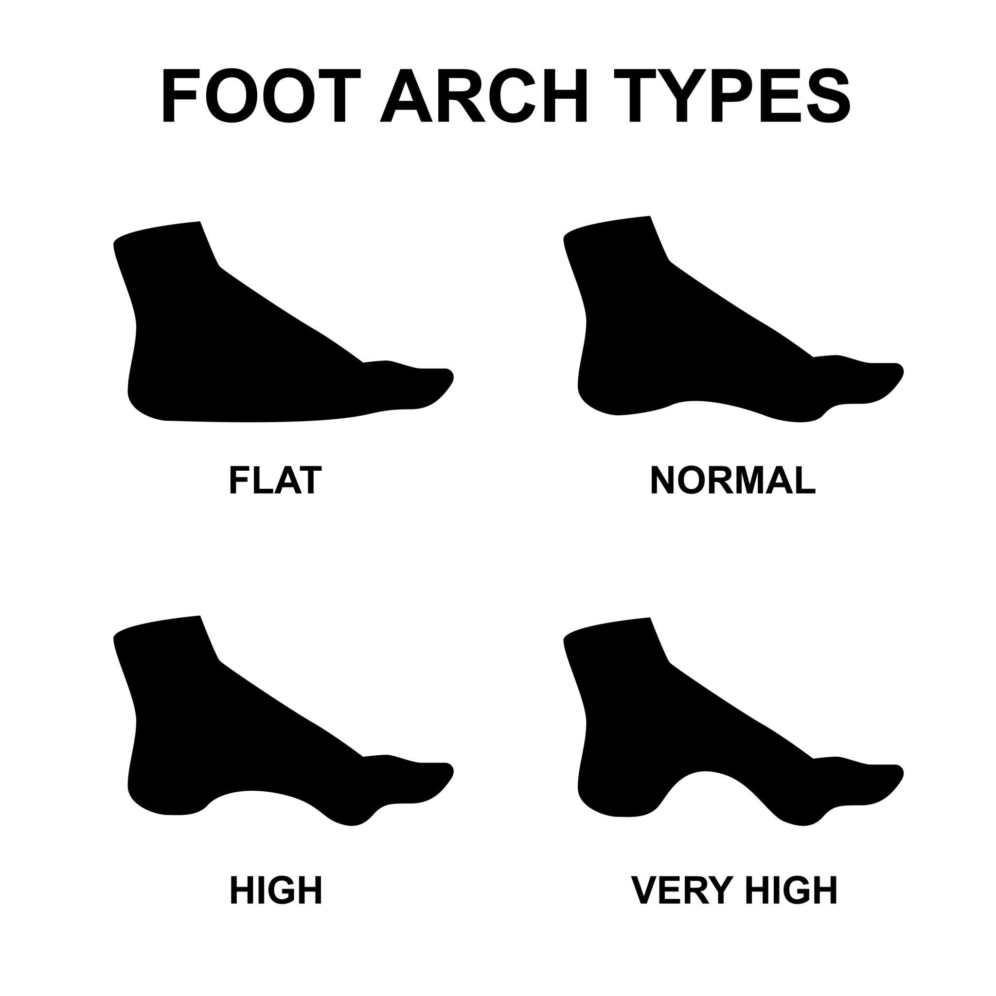
Foot arch height can generally be categorised into three main types:
1. Normal Arch (Neutral Foot)
A normal arch provides optimal shock absorption and weight distribution. This foot type allows for efficient movement and minimal stress on the lower body (Kappel-Bargas et al., 1998).
2. Flat Foot (Pes Planus)
A flat foot lacks proper arch structure, causing excessive pronation. This foot type is associated with overuse injuries and joint instability (Buldt et al., 2018).
3. High-Arched Foot (Pes Cavus)
A high-arched foot has an exaggerated arch that reduces ground contact, leading to increased pressure on the heel and forefoot. This condition alters gait mechanics and often causes rigidity in the foot (Coughlin et al., 2016).
Differences Between Each Arch Type
Each type of foot arch has unique structural and functional characteristics that impact movement and weight distribution. Below is a breakdown of the differences:
1. Normal Arch (Neutral Foot):
This foot type has a balanced structure with even weight distribution across the heel, midfoot, and forefoot.
• The medial longitudinal arch maintains a healthy shape, allowing for efficient shock absorption and force transfer during walking and running.
• Individuals with a normal arch typically experience minimal stress on the joints, leading to lower risk of overuse injuries.
2. Flat Foot (Pes Planus):
A low or collapsed arch leads to excessive inward rolling of the foot, known as overpronation.
• This increases stress on the ankles, knees, and hips, as the body’s alignment is altered.
• Flat feet are associated with higher risks of plantar fasciitis, shin splints, and knee pain due to inefficient weight distribution.
3. High-Arched Foot (Pes Cavus):
This foot type features an exaggerated arch height, which reduces the contact surface with the ground.
• The high arch leads to excessive weight being placed on the heel and forefoot, causing pressure points and discomfort.
• Since this foot type is often more rigid, it lacks natural shock absorption, increasing the risk of ankle instability, stress fractures, and lower back pain.
Key Functional Differences:
• Flat feet lead to excessive mobility and stress on the joints, while high arches cause reduced shock absorption and rigidity.
• A normal arch allows for balanced weight distribution and optimal movement mechanics.
• People with high arches tend to supinate (roll outward) excessively, whereas those with flat feet overpronate (roll inward).
A high-arched foot differs significantly in function compared to the other two types. Unlike a neutral or flat foot, which provides more even force distribution, pes cavus leads to inefficient weight-bearing and greater stress on surrounding joints (Burns et al., 2005).
High-Arched Foot
A high-arched foot is less common than a flat foot and is typically more rigid. This foot type lacks the ability to absorb shock efficiently and places excessive pressure on the heel and forefoot (McPoil et al., 2009). The limited flexibility in a high-arched foot reduces the body's ability to adapt to uneven surfaces, increasing the risk of injuries.
Effects on the Lower Leg and Body
A high-arched foot leads to several compensatory changes in the body:
1. Increased Load on the Heel and Forefoot
- Due to reduced surface area contact, more stress is placed on the calcaneus (heel bone) and metatarsals (Kelly et al., 2014).
2. Ankle Instability and Sprains
- High arches often cause excessive supination, leading to unstable lateral movement and a greater risk of ankle sprains (Rao et al., 2011).
3. Knee and Hip Misalignment
- The rigid structure of a high-arched foot alters the tibia's rotation, affecting knee tracking and hip positioning (Neumann, 2017).
4. Lower Back Pain
- Altered foot mechanics can contribute to compensatory movement patterns, placing excessive stress on the lumbar spine (Buldt et al., 2018).
Causes of a High-Arched Foot
Several factors contribute to developing a high-arched foot:
- Apparently Genetics– Many cases of pes cavus are inherited (Burns et al., 2005).
- Neurological Conditions – Disorders like Charcot-Marie-Tooth disease or cerebral palsy can cause muscle imbalances, leading to high arches (Coughlin et al., 2016).
- Muscle Imbalances – Weakness in certain foot and leg muscles may contribute to increased arch height (Rao et al., 2011).
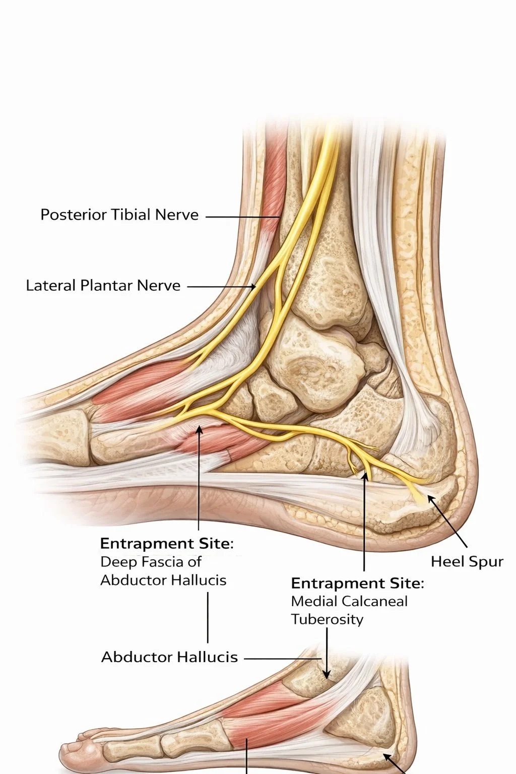
Relationship of Bones to Body Mechanics
The structure of the foot affects the alignment of the entire body. In a high-arched foot:
- The calcaneus is often inverted, dorsiflexed and externally rotated affecting ankle motion as this is also rotated externally.
- The tibia externally rotates , altering knee and hip positioning.
- Pelvic alignment is affected learning to a posterior tilted pelvis and strain on the lumbar spine and compensatory spinal movements (McPoil et al., 2009).
These changes influence gait mechanics, making efficient movement more difficult.
Movement Restrictions in a High-Arched Foot
- Limited Pronation: Reduces shock absorption.
- Restricted Ankle Dorsiflexion: Increases strain on the Achilles tendon.
- Stiff Midfoot: Reduces adaptability to uneven surfaces.
- Increased Load on Metatarsal Heads: Contributes to forefoot pain and stress fractures (Kelly et al., 2014).
Long-Term Effects on Foot Function
Without intervention, a high-arched foot can lead to:
- Chronic pain (heel, forefoot, knees, and lower back).
- Foot deformities such as claw toes and hammertoes.
- Plantar fasciitis and Achilles tendonitis.
- Increased risk of stress fractures due to high-pressure points (Rao et al., 2011).
What You Can Do to Improve Function
1. Strengthening and Mobility Exercises
- Foot arch mobility drills to improve flexibility.
- Calf stretching to counteract tightness.
- Intrinsic foot strengthening (e.g., calf raises) to enhance support.
2. Proper Footwear
- Shoes with good arch support and cushioning can help distribute pressure more evenly.
- Avoid rigid footwear that limits natural foot motion.
3. Orthotics and Insoles ( I don’t this so but research says otherwise)
- Custom orthotics can help reduce excessive supination and redistribute weight (Buldt et al., 2018).
4. Manual Therapy and Gait Retraining
- Movement therapy can help improve movement mechanics.
- Massage and myofascial release can reduce stiffness and pain.
Conclusion
A high-arched foot presents unique biomechanical challenges that can lead to long-term musculoskeletal issues. Understanding its effects and implementing corrective strategies can help reduce pain, improve function, and prevent further complications. If you experience discomfort related to high arches, seeking professional advice for assessment and treatment is highly recommended.
References
• Buldt, A. K., Allan, J. J., Landorf, K. B., Menz, H. B., Hill, K. D. and Vicenzino, B., 2018. Foot posture is associated with kinematics of the lower but not upper limb during walking and running. Gait & Posture, 62, pp.230-238.
• Burns, J., Crosbie, J., Hunt, A. and Ouvrier, R., 2005. The effect of pes cavus on foot pain and plantar pressure.Clinical Biomechanics, 20(9), pp.877-882.
• Coughlin, M. J., Mann, R. A. and Saltzman, C. L., 2016. Mann’s Surgery of the Foot and Ankle. 9th ed. Philadelphia: Elsevier.
• Kelly, L. A., Girard, O. and Racinais, S., 2014. Effects of arch structure and footwear on ankle and knee mechanics in running. Medicine and Science in Sports and Exercise, 46(5), pp.883-891.
• Kappel-Bargas, A., Woolf, R. D., Cornwall, M. W. and McPoil, T. G., 1998. The influence of foot structure on plantar pressure variations during walking. Journal of the American Podiatric Medical Association, 88(6), pp.285-291.
• McPoil, T. G., Cornwall, M. W., Vicenzino, B. and Teyhen, D. S., 2009. Foot orthoses and gait: The role of transverse plane kinetics. Journal of Orthopaedic & Sports Physical Therapy, 39(10), pp.733-740.
• Neumann, D. A., 2017. Kinesiology of the Musculoskeletal System: Foundations for Rehabilitation. 3rd ed. St. Louis: Elsevier.
• Rao, S., Saltzman, C. L. and Yack, H. J., 2011. Relationship of foot structure and function to lower extremity injury: A review of literature. Clinical Biomechanics, 26(5), pp.547-555.