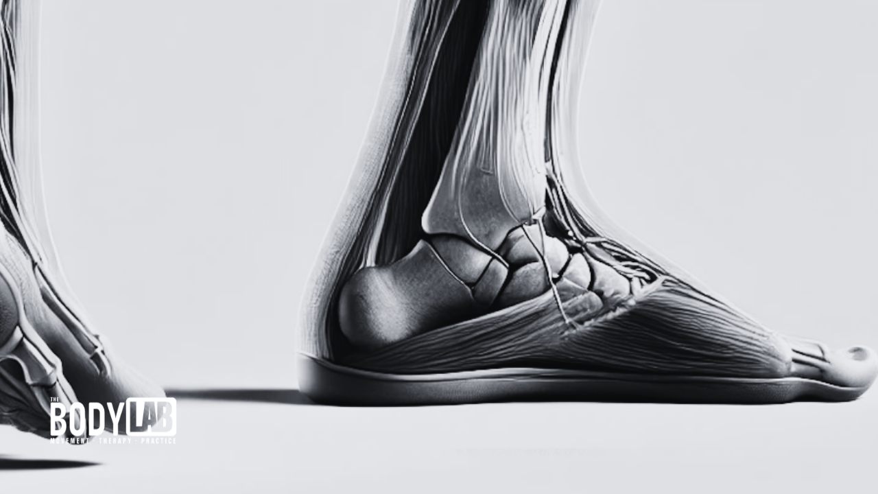
Common Issues with Pronation and Their Impact on Joint Mechanics
What is pronation
Pronation is a key component of gait, enabling the lower limb to absorb shock, adapt to uneven surfaces, and transition efficiently from one phase of movement to the next. However, when optimal pronation mechanics are not achieved, it can lead to a host of issues that affect the entire kinetic chain, from the foot to the pelvis. Understanding how each joint is impacted by dysfunctional pronation, and the downstream consequences of such dysfunction, is crucial for healthcare professionals working in movement therapy, rehabilitation, and sports performance.
In this article, we will explore common issues that arise when pronation mechanics fail, focusing on the rearfoot, forefoot, talocrural (ankle) and subtalar joints, knee, hip, and pelvis. These issues are rooted in both underpronation (supination) and overpronation, and each joint’s response to such dysfunction can cause an array of musculoskeletal problems.
Defining Pronation: History, Medical Terminology, and Aetiology
Pronation, from a medical perspective, refers to a triplanar movement that occurs in the four major sections of the foot: the talocrural (ankle) joint, rearfoot, forefoot, and digits. It’s important to note that the entire body undergoes pronation, shaping itself to eccentrically load muscles in order to absorb the forces generated from heel strike through the loading phase of gait. However, this discussion will focus solely on foot mechanics.
The foot moves in three dimensions, rotating around three axes within the sagittal, frontal, and transverse planes. These movements include dorsiflexion and plantarflexion in the sagittal plane, eversion and inversion in the frontal plane, and internal or external rotation in the transverse plane. While some research papers use the terms “abduction” and “adduction” to describe foot motions, I avoid these terms for greater clarity. These combined motions allow the foot to adapt to uneven surfaces and absorb shock efficiently during gait.
The term “pronation” originates from the Latin word pronatus, meaning “bent forward.” The biomechanical importance of pronation was extensively studied in the mid-20th century by anatomists like John Hicks, who described the Windlass Mechanism and its role in foot stability during gait (Hicks, 1954).
Pronation is specifically defined as the rearfoot undergoing plantarflexion, eversion, and internal rotation, while the forefoot simultaneously dorsiflexes, inverts, and externally rotates, with the talocrural (ankle) joint in dorsiflexion.
Dysfunction in pronation can manifest as either overpronation—where the foot rolls excessively inward—or underpronation, where the foot remains overly rigid, limiting its ability to absorb shock. Both forms of dysfunction can disrupt the natural biomechanics of gait, leading to compensatory movement patterns that negatively impact the joints of the lower limb.
Rearfoot Mechanics and Common Pronation Issues
The rearfoot, consisting primarily of the calcaneus and navicular bones, plays a critical role in initiating pronation during the loading phase of gait, which begins after the rearfoot makes contact with the ground through initial contact (heel strike). Pronation is typically defined as the moment the medial aspect of the foot—particularly the first metatarsal joint—engages the ground during the loading phase. Dysfunctional mechanics at the rearfoot can lead to common issues, such as overpronation or underpronation.
Overpronation:
In overpronation, the calcaneus experiences exaggerated motion across all three planes (sagittal, frontal, and transverse). This exaggerated motion can result from improper positioning during heel strike. For effective pronation mechanics, the calcaneus should display an optimal heel strike pattern, meaning that before the foot transitions into plantar flexion, eversion, and internal rotation during pronation, it must first land in dorsiflexion, inversion, and external rotation. Overpronation occurs when the foot remains in the pronated position for too long, failing to achieve neutral positioning by midstance and continuing to delay transition into the supination phase (from midstance to toe-off). This prolonged pronation compromises the rearfoot’s ability to resupinate, leaving the foot flexible during a period when it should be acting as a rigid lever for effective propulsion. Over time, this excessive motion places stress on the tibialis posterior tendon, contributing to posterior tibial tendon dysfunction (PTTD). Research by Kulig et al. (2009) links PTTD with chronic overpronation, which can lead to flatfoot deformity and ongoing pain.
Underpronation (Supination):
In underpronation, the rearfoot experiences insufficient pronation or no meaningful engagement of the pronation mechanics during the loading phase of gait. The foot remains more neutral or in a supinated position—characterized by dorsiflexion, inversion, and external rotation—throughout the loading phase, limiting its ability to absorb shock. When the foot fails to pronate properly, the dissipation of ground reaction forces falls onto other structures, increasing the load on the lateral components of the foot and ankle. This imbalance often leads to conditions such as peroneal tendonitis. Razeghi and Batt (2000) suggest that underpronation also heightens the risk of stress fractures, especially in the lateral foot and leg, due to the lack of adaptability to ground forces.
Forefoot Mechanics and Dysfunction
The forefoot must adapt to the rearfoot’s motion to maintain balance and alignment during gait. It must also oppose the rear foot’s movements to allow the foot to become a flexible and dynamic stabiliser, capable of adapting to different surfaces during walking. This adaptability is achieved through the foot’s three distinct arches, which form a tripod structure. When forefoot mechanics are disrupted due to abnormal or delayed pronation, it can lead to a variety of foot pathologies. These issues often arise from insufficient time within the gait cycle to allow all joints to fully express their range of motion. Gait, therefore, is a finely-tuned balance of timing that enables each joint to work harmoniously within every stride.
Overpronation:
Excessive pronation at the rearfoot forces the forefoot into compensatory dorsiflexion, inversion, and external rotation, which places strain on the first metatarsophalangeal (MTP) joint. This compensation can result in the formation of hallux valgus (bunions) and increased stress on the plantar fascia, as the foot tries to generate more motion potential. Additionally, the foot may exhibit external rotation, causing it to “turn out” in an attempt to increase its range of motion. According to Sangeorzan and Mosca (1993), prolonged overpronation leads to structural deformities such as metatarsus primus elevatus, where the first metatarsal becomes elevated. This deformity contributes to Windlass Mechanism dysfunction, reducing push-off efficiency during gait.
Underpronation:
When the forefoot does not sufficiently pronate to compensate for rearfoot motion, it increases pressure and compression on the lateral forefoot. This can result in metatarsalgia, particularly in runners or athletes who place heavy loads on the forefoot. Studies by Cornwall and McPoil (1999) suggest that individuals with underpronation are at a higher risk for metatarsal stress fractures and Morton’s neuroma due to poor load distribution across the forefoot. It is important to note that the fourth and fifth metatarsals are not designed to bear excessive loads, and undue force in this region can lead to stress fractures.

Talocrural and Subtalar Joint Dysfunction
The talocrural and subtalar joints work in tandem during pronation, ensuring smooth transitions through the gait cycle by transferring ground reaction forces up the limb. The position of the subtalar joint directly influences the motion of the talocrural (ankle) joint. The talus, situated between the distal ends of the tibia and fibula, primarily functions in the sagittal plane but exhibits frontal and transverse planes via the subtalar joint, specifically through the actions of the rearfoot. Dysfunction at either of these joints can have significant effects on overall gait mechanics.
Talocrural Joint Dysfunction:
The talocrural joint is responsible for most of the dorsiflexion and plantarflexion at the ankle. However, dysfunction at this joint, such as excessive frontal plane motion (eversion), can limit dorsiflexion and cause compensatory movements in other joints. When the talocrural joint exhibits excessive inward rolling over the subtalar joint, the result is inefficient transfer of force and abnormal loading patterns. According to Whittle (2014), restricted dorsiflexion due to faulty pronation mechanics can lead to compensatory knee hyperextension or contribute to Achilles tendinopathy, as the Achilles tendon is subjected to increased strain during the push-off phase.
When the talocrural joint is unable to dorsiflex due to an inverted position over the subtalar joint, force is inadequately transferred up the limb. This dysfunction prevents proper loading of muscles and may impair the body’s preparation for supination in the later stages of gait.
Subtalar Joint Dysfunction:
The subtalar joint plays a critical role in controlling inversion and eversion of the foot, particularly in the rearfoot. In cases of overpronation, the subtalar joint remains excessively plantarflexed, everted, and internally rotated, which hinders the foot’s ability to resupinate in preparation for push-off. This prolonged eversion compromises the foot’s structural stability and can lead to conditions such as tibialis posterior tendonitis or even progressive flatfoot deformity (Nester et al., 2002).
On the other hand, underpronation limits the subtalar joint’s ability to properly adapt to ground reaction forces, which increases the risk of lateral ankle sprains. In this scenario, the foot remains overly inverted and rigid, reducing shock absorption and increasing stress on the lateral structures of the foot and ankle.
Knee Mechanics and Pronation Issues:
Pronation is closely linked to knee function, as it directly influences tibial rotation and knee alignment. Dysfunctional pronation, especially excessive pronation, significantly impacts knee health. This can cause the knee to remain excessively flexed and externally rotated during the stance phase of gait, limiting its ability to reverse these movements efficiently. The relationship between the knee ligaments and hip joint is often underestimated, particularly in how femoral rotation affects ligament health.
Overpronation:
In cases of overpronation, there is prolonged and increased internal rotation of the tibia, placing excessive strain on the knee joint, especially in the medial compartment, as the tibia continues to rotate, causing compensatory external rotation of the knee. This increased tibial and femoral rotation alters patellofemoral mechanics, often leading to conditions like patellofemoral pain syndrome (PFPS) and medial knee osteoarthritis. According to research by Powers (2010), overpronation significantly contributes to abnormal knee kinematics. Women are particularly susceptible due to their wider pelvic anatomy and increased Q-angle, which further exacerbates knee dysfunctions.
Underpronation:
Conversely, underpronation limits tibial rotation and restricts femoral movement and knee flexion over the lateral compartment of the foot, which can result in increased strain on the lateral knee structures. With inadequate pronation, the foot’s shock absorption capacity diminishes, transferring excessive ground reaction forces to the knee. This increases the risk of developing iliotibial band syndrome (ITBS), particularly in runners. In these individuals, the lack of proper shock absorption further stresses the IT band, aggravating the condition (Nicol et al., 2010).
Hip Mechanics and Pronation-Related Issues
The hip joint, like the knee, is significantly influenced by pronation mechanics, which are essential for maintaining long-term joint health. Dysfunctional pronation can lead to compensatory movements that increase the risk of hip instability and pain. The pelvis and hip joint are intrinsically connected, working together to dissipate forces generated during heel strike and the loading phase of gait. Proper pronation mechanics ensure efficient force distribution, reducing strain on the hip and pelvis while promoting stability throughout the gait cycle.
Overpronation:
Excessive pronation causes increased internal tibial and femoral rotation, which leads to excessive internal rotation at the hip joint. Over time, this can cause hip instability, femoroacetabular impingement (FAI), and labral tears. Levinger and Gilleard (2010) found that excessive pronation is linked to hip adduction and internal rotation, which creates excessive stress on the hip joint’s ligaments and structures. Overpronation may also affect the pelvis, increasing anterior pelvic tilt, which places further stress on the lower back and hips. This overloading of the hip can lead to gluteal tendinopathy and other hip-related conditions, including bursitis.
Additionally, as the foot remains excessively pronated, the body’s center of mass shifts medially, causing increased demand on the hip abductors to stabilise the pelvis during walking. This prolonged demand can lead to overuse injuries in the hip musculature, particularly the gluteus medius and minimus, which are responsible for maintaining pelvic stability. A weakened gluteal complex results in a “Trendelenburg gait,” where the pelvis drops on the contralateral side during single-leg stance.
Underpronation:
Underpronation, or insufficient pronation, restricts the foot’s ability to accommodate ground forces and absorb shock, which can lead to compensatory external rotation at the hip joint. When the foot fails to pronate adequately, it places increased stress on the lateral aspect of the hip and contributes to issues such as greater trochanteric pain syndrome (GTPS). The gluteus medius and other lateral hip muscles are forced to compensate for the lack of shock absorption and flexibility in the lower limb. As a result, underpronators are prone to developing hip and pelvic pain due to the increased lateral forces.
Underpronation also limits the natural internal rotation of the femur that typically occurs during the stance phase of gait. This lack of internal rotation can place strain on the hip joint and alter the natural movement patterns of the pelvis. Restricted femoral internal rotation may exacerbate hip impingement and contribute to long-term joint degeneration.
Conclusion: Pronation and Their Impact on Joint Mechanics
The mechanics of pronation play an essential role in ensuring optimal gait and overall lower limb function. Proper pronation allows the foot to absorb shock, adapt to different surfaces, and distribute forces evenly throughout the kinetic chain. However, when dysfunctional pronation occurs—whether it is overpronation or underpronation—the entire system of joints from the foot to the pelvis is disrupted. This disruption can result in compensatory movement patterns that increase the risk of injury, pain, and chronic musculoskeletal issues.
Overpronation leads to excessive internal tibial rotation, stressing the medial compartments of the knee and hip, which can result in knee osteoarthritis, patellofemoral pain syndrome (PFPS), and conditions like posterior tibial tendon dysfunction (PTTD). Prolonged overpronation also affects the forefoot, placing strain on the first metatarsophalangeal (MTP) joint and potentially leading to deformities like hallux valgus. On the other hand, underpronation limits the foot’s shock-absorbing capacity, which can result in increased stress on the lateral structures of the foot and ankle, leading to conditions like peroneal tendonitis, metatarsal stress fractures, and iliotibial band syndrome (ITBS).
Dysfunctional pronation, whether excessive or insufficient, has far-reaching consequences that extend beyond the foot. From increased knee and hip instability to compensatory pelvic tilt, the entire lower kinetic chain is affected. It is crucial for healthcare professionals, particularly those involved in rehabilitation, movement therapy, and sports performance, to understand the mechanics of pronation and its downstream effects on joint health.
By addressing these dysfunctions early, whether through corrective exercises, orthotics, or targeted interventions, clinicians can help patients achieve more efficient movement patterns, reduce their risk of injury, and improve long-term joint health.
References
• Cornwall, M. W., & McPoil, T. G. (1999). Footwear and foot orthoses. In M. W. Cornwall & T. G. McPoil (Eds.), Clinical biomechanics of the lower extremities (pp. 143-168). Elsevier Health Sciences.
• Hicks, J. H. (1954). The mechanics of the foot: II. The plantar aponeurosis and the arch. Journal of Anatomy, 88(1), 25-30.
• Kulig, K., Reischl, S. F., Pomrantz, A. B., Burnfield, J. M., Mais-Requejo, S., Thordarson, D. B., & Smith, R. W. (2009). Nonsurgical management of posterior tibial tendon dysfunction with orthoses and resistive exercise: A randomized controlled trial. Physical Therapy, 89(1), 26-37.
• Levinger, P., & Gilleard, W. (2010). The relationship between foot pronation and hip mechanics in individuals with patellofemoral pain syndrome. Journal of Orthopaedic & Sports Physical Therapy, 40(2), 42-51.
• Nester, C. J., Findlow, A. H., & Bowker, P. (2002). Mechanisms used to accommodate foot structure and the orthosis design process. Gait & Posture, 15(1), 48-55.
• Nicol, C., Komi, P. V., & Marconnet, P. (2010). Fatigue effects of marathon running on neuromuscular performance: Changes in force, integrated electromyographic activity, and endurance. European Journal of Applied Physiology, 55(1), 71-79.
• Powers, C. M. (2010). The influence of abnormal hip mechanics on knee injury: A biomechanical perspective. Journal of Orthopaedic & Sports Physical Therapy, 40(2), 42-51.
• Razeghi, M., & Batt, M. E. (2000). Biomechanical analysis of the effect of orthotic shoe inserts: A review of the literature. British Journal of Sports Medicine, 34(2), 89-96.
• Sangeorzan, B. J., & Mosca, V. (1993). Biomechanics and pathophysiology of the adult acquired flatfoot. Foot & Ankle, 14(6), 357-361.
• Whittle, M. W. (2014). Gait analysis: An introduction. Elsevier Health Sciences.
